


COIMBATORE RETINA MEET - March 18, 2026
COIMBATORE RETINA MEET
## Minutes of Meeting
*Date:* March 18, 2026
*Time:* 7:00 PM onwards
*Venue:* Zone by the Park, Coimbatore
—–
## MEETING OVERVIEW
The Coimbatore Retina Meet convened regional vitreoretinal specialists and ophthalmologists to explore cutting-edge advancements in retinal imaging technology and artificial intelligence applications in ophthalmology. The scientific session featured dual keynote lectures by Professor Dr. J. Peter Campbell from Oregon Health & Science University, addressing ultra-widefield optical coherence tomography and AI-driven diagnostics for retinopathy of prematurity. The program included five compelling case presentations addressing challenging clinical scenarios in retinal vascular disease, retinopathy of prematurity, and ocular toxicology.
—–
## PROGRAM PROCEEDINGS
### 1. WELCOME AND CONTEXT SETTING (7:00 PM)
*Presenter:* Dr. George Manayath
Dr. Manayath inaugurated the proceedings by welcoming all delegates to this educational forum focused on technological innovation in vitreoretinal practice. The introduction provided context on the transformative impact of ultra-widefield imaging and artificial intelligence in reshaping diagnostic paradigms and treatment decision-making in contemporary retinal surgery. Dr. Manayath emphasized the importance of adopting evidence-based imaging technologies and understanding AI integration to optimize clinical outcomes, particularly in underserved populations where screening efficiency is paramount. The session objectives were outlined, highlighting knowledge translation from international expertise to regional practice settings.
### 2. INTRODUCTION OF GUEST SPEAKER (7:10 PM)
*Presenter:* Dr. Parag Shah
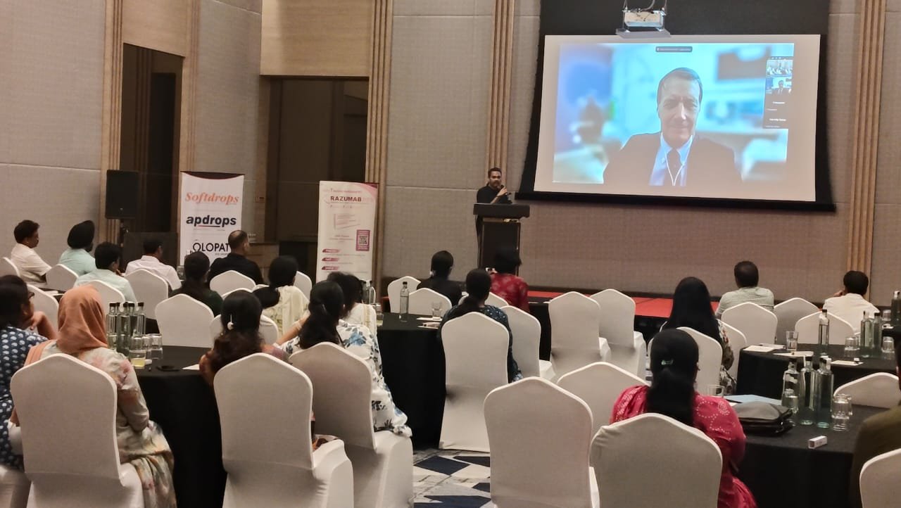
Dr. Parag Shah introduced the distinguished international guest lecturer and assumed the role of session chair. The introduction highlighted Professor Dr. J. Peter Campbell’s credentials as Professor of Ophthalmology at the School of Medicine, Oregon Health & Science University (OHSU), one of North America’s premier academic medical centers. Dr. Shah acknowledged Professor Campbell’s seminal contributions to pediatric retinal imaging, artificial intelligence development for retinopathy of prematurity screening, and advancement of ultra-widefield OCT technology. The introduction noted Professor Campbell’s extensive publication record in high-impact journals and his leadership in translating AI research into clinical practice. Dr. Shah expressed gratitude for Professor Campbell’s willingness to share international expertise with the South Indian ophthalmology community.
—–
## KEYNOTE LECTURES
### 3. GUEST LECTURE PART 1: “ULTRA-WIDEFIELD OCT IN VITREORETINAL SURGERY”
*Speaker:* Prof. Dr. J. Peter Campbell, M.D., M.P.H.
*Designation:* Professor of Ophthalmology, Oregon Health & Science University
Professor Campbell delivered the first segment of his lecture, providing comprehensive insights into ultra-widefield optical coherence tomography and its surgical applications:
#### Technological Overview
– *Evolution of OCT Technology:* Progression from time-domain to spectral-domain to swept-source OCT, and the development of ultra-widefield capabilities extending beyond the traditional posterior pole
– *Ultra-Widefield OCT Platforms:* Comparative analysis of available systems, technical specifications including scan width (typically 100-200 degrees), resolution parameters, and acquisition speed
– *Imaging Principles:* Swept-source technology utilizing longer wavelength infrared light (1050 nm) enabling deeper penetration through media opacities and enhanced visualization of choroidal structures
– *Multimodal Integration:* Combination of ultra-widefield OCT with color fundus photography, autofluorescence, and fluorescein angiography for comprehensive retinal assessment
#### Clinical Applications in Vitreoretinal Surgery
*Preoperative Planning:*
– *Peripheral Pathology Detection:* Identification of occult peripheral retinal breaks, lattice degeneration, retinoschisis, and peripheral neovascularization not visible on conventional posterior pole OCT
– *Diabetic Retinopathy Assessment:* Comprehensive evaluation of peripheral ischemia, neovascular complexes, and fibrovascular proliferation extending beyond the vascular arcades
– *Retinal Detachment Evaluation:* Precise localization of retinal breaks, assessment of proliferative vitreoretinopathy distribution, and identification of subclinical detachment extensions
– *Surgical Risk Stratification:* Enhanced ability to predict surgical complexity and plan appropriate interventions based on peripheral pathology extent
*Intraoperative Utilization:*
– *Real-time Surgical Guidance:* Integration of intraoperative OCT systems providing immediate feedback on membrane peeling completeness, internal limiting membrane removal, and retinal apposition
– *Vitrectomy Optimization:* Confirmation of posterior vitreous detachment induction and identification of residual vitreous adhesions to peripheral retina
– *Retinotomy Assessment:* Evaluation of drainage retinotomy edges and confirmation of subretinal fluid drainage in complex retinal detachment repair
*Postoperative Monitoring:*
– *Anatomical Outcome Assessment:* Comprehensive evaluation of retinal reattachment, foveal contour restoration, and detection of subclinical recurrent detachment
– *Complication Detection:* Early identification of epiretinal membrane formation, macular hole edges, cystoid macular edema, and peripheral retinal pathology
– *Long-term Surveillance:* Monitoring for late complications including proliferative vitreoretinopathy recurrence and peripheral retinal breaks
#### Research Applications and Future Directions
– *Quantitative Analysis:* Automated measurement of retinal layer thickness across the entire retina, choroidal thickness mapping, and volumetric assessments
– *AI Integration:* Machine learning algorithms for automated pathology detection in ultra-widefield OCT scans
– *Pediatric Applications:* Particular utility in retinopathy of prematurity evaluation and pediatric retinal dystrophy assessment where peripheral changes are prominent
– *Screening Programs:* Potential for ultra-widefield OCT in diabetic retinopathy and sickle cell retinopathy screening initiatives
The lecture incorporated extensive imaging examples from OHSU’s clinical experience, demonstrating practical applications across diverse vitreoretinal pathologies.
### 4. GUEST LECTURE PART 2: “AI FOR ROP (RETINOPATHY OF PREMATURITY)”
*Speaker:* Prof. Dr. J. Peter Campbell, M.D., M.P.H.
Professor Campbell’s second lecture segment focused on artificial intelligence applications in retinopathy of prematurity diagnosis and management:
#### ROP Burden and Screening Challenges
– *Global Epidemiology:* ROP as a leading cause of childhood blindness worldwide, with particular burden in middle-income countries experiencing rapid expansion of neonatal intensive care without proportional ophthalmology expertise
– *Screening Requirements:* Need for serial examinations of at-risk premature infants, resource intensity, and shortage of trained ROP specialists
– *Interobserver Variability:* Documented inconsistencies in ROP classification among experienced ophthalmologists, highlighting the need for objective diagnostic support
– *Access Barriers:* Geographic mismatch between NICU locations and ROP specialist availability, particularly in rural and underserved regions
#### AI System Development and Validation
*Training Methodology:*
– *Image Dataset Curation:* Description of large-scale retinal image databases used for AI training, including the i-ROP cohort study with thousands of annotated wide-angle retinal photographs
– *Deep Learning Architecture:* Convolutional neural networks designed specifically for retinal image analysis, transfer learning from general image recognition models
– *Ground Truth Establishment:* Expert consensus grading protocols and resolution of intergrader disagreements to create reliable reference standards
– *Data Augmentation:* Techniques to enhance model robustness including rotation, brightness adjustment, and synthetic image generation
*Performance Metrics:*
– *Diagnostic Accuracy:* Presentation of sensitivity and specificity data for AI systems detecting plus disease, pre-plus disease, and treatment-requiring ROP
– *Comparison with Human Experts:* Head-to-head performance evaluation demonstrating AI systems achieving expert-level or superior diagnostic accuracy
– *External Validation:* Testing on independent datasets from different geographic regions and imaging platforms to confirm generalizability
– *Real-world Performance:* Results from prospective implementation studies in clinical NICU settings
#### Clinical Implementation
*Workflow Integration:*
– *Image Acquisition:* Standardized protocols for wide-angle retinal photography in premature infants using commercially available systems
– *AI Analysis Pipeline:* Automated upload, processing, and diagnostic output generation within minutes of image capture
– *Decision Support:* AI-generated risk scores, zone classification, stage determination, and treatment recommendations
– *Human Oversight:* Framework for ophthalmologist review of AI outputs, with particular attention to borderline cases and AI uncertainty flags
*Telemedicine Applications:*
– *Remote Screening:* AI-enabled ROP screening in facilities without on-site ophthalmologists
– *Triage Systems:* Prioritization of high-risk infants requiring urgent specialist evaluation
– *Quality Assurance:* Automated image quality assessment rejecting suboptimal photographs before AI analysis
– *Reading Center Models:* Centralized expert review systems augmented by AI pre-screening
#### Regulatory and Ethical Considerations
– *FDA Approval Status:* Discussion of regulatory pathways for AI medical devices and current approved systems for ROP screening
– *Liability Framework:* Medico-legal considerations for AI-assisted diagnosis and recommendations for appropriate documentation
– *Algorithmic Bias:* Potential for AI performance variations across different populations and importance of diverse training datasets
– *Physician-AI Collaboration:* Optimal models for integrating AI recommendations with clinical judgment
#### Future Directions and Innovation
– *Predictive Modeling:* AI systems forecasting ROP progression and individualizing examination frequency
– *Treatment Response Prediction:* Algorithms identifying infants at high risk for treatment failure or requiring augmented therapy
– *Multimodal AI:* Integration of clinical data (gestational age, birth weight, oxygen exposure) with imaging for enhanced prediction
– *Global Implementation:* Strategies for deploying AI-ROP systems in resource-limited settings including offline capabilities and smartphone-based solutions
Professor Campbell concluded with case examples demonstrating AI system performance in challenging diagnostic scenarios and discussed OHSU’s ongoing research initiatives in this field.
—–
## CASE PRESENTATIONS
### 5. NON-INVASIVE RETINAL DYE ANGIOGRAPHY
*Presenter:* Dr. Saurab Kapase, The Eye Foundation
This presentation examined non-invasive alternatives to traditional fluorescein and indocyanine green angiography. Discussion included:
– *Technological Background:*
– *OCT Angiography (OCTA):* Principles of motion contrast imaging to visualize blood flow without dye injection
– *Ultra-widefield Autofluorescence:* Lipofuscin and melanin distribution patterns providing vascular and pathology information
– *Adaptive Optics Imaging:* Direct visualization of individual capillaries and photoreceptors
– *Clinical Applications:*
– *Diabetic Retinopathy:* OCTA detection of capillary non-perfusion, microaneurysms, and neovascularization without dye-related artifacts
– *Age-related Macular Degeneration:* Choroidal neovascularization characterization, flow patterns, and treatment monitoring
– *Retinal Vascular Occlusions:* Quantification of ischemic areas and collateral vessel assessment
– *Uveitis Evaluation:* Non-invasive vasculitis assessment and macular edema characterization
– *Advantages Over Traditional Angiography:*
– *Patient Safety:* Elimination of allergic reactions, nausea, and rare anaphylaxis risk
– *Repeatability:* Ability to perform multiple examinations in single visit without dye washout delays
– *Depth Resolution:* Layer-by-layer vascular visualization (superficial, intermediate, deep capillary plexuses)
– *Quantitative Analysis:* Automated measurement of foveal avascular zone, vessel density, and flow parameters
– *Limitations and Challenges:*
– *Field of View:* Current OCTA systems limited to posterior pole versus widefield capability of traditional angiography
– *Motion Artifacts:* Patient cooperation requirements and image quality degradation with eye movement
– *Media Opacity Sensitivity:* Reduced performance with cataract, vitreous hemorrhage, or corneal pathology
– *Learning Curve:* Interpretation differences from traditional angiography requiring expertise development
– *Comparative Case Examples:* Side-by-side presentations of traditional angiography versus OCTA findings in identical patients, demonstrating concordance and unique information from each modality
– *Future Directions:*
– Widefield OCTA development
– Enhanced depth imaging for choroidal vasculature
– AI-assisted OCTA interpretation and quantification
– Integration with multimodal imaging platforms
– *Clinical Decision Making:* Guidelines for selecting appropriate imaging modality based on clinical question, patient factors, and available technology
### 6. DOUBLE EDGED SWORD: LOST TO FOLLOW-UP AFTER ANTI-VEGF INJECTION IN ROP
*Presenter:* Dr. Bidisha P, Aravind Eye Hospital
This presentation addressed the critical challenge of treatment adherence in retinopathy of prematurity management. Content included:
– *Background and Context:*
– *Anti-VEGF as ROP Treatment:* Paradigm shift from laser photocoagulation to intravitreal bevacizumab/ranibizumab for Type 1 ROP, particularly Zone I disease
– *Theoretical Advantages:* Preservation of peripheral retinal development, potential for vascularization completion, reversibility compared to destructive laser
– *Critical Follow-up Requirements:* Need for intensive monitoring given late reactivation risk, extended vulnerable period compared to laser treatment
– *Case Presentation:*
– *Patient Demographics:* Presentation of specific case(s) involving premature infants treated with anti-VEGF who subsequently became lost to follow-up
– *Treatment Details:* Initial ROP staging, decision-making for anti-VEGF over laser, injection technique, and initial response
– *Follow-up Loss:* Timeline of missed appointments, attempts at patient contact, and eventual loss of contact with family
– *Late Presentation:* Description of eventual return weeks to months later with advanced disease progression
– *Consequences of Treatment Abandonment:*
– *Late Reactivation Phenomenon:* ROP progression occurring months after initial anti-VEGF response, often beyond typical laser treatment window
– *Severity at Return:* Stage 4 or 5 ROP, tractional retinal detachment, or end-stage disease requiring complex surgery or resulting in blindness
– *Salvage Options:* Limited therapeutic alternatives (vitrectomy in stage 4-5, often with poor prognosis)
– *Visual Outcomes:* Poor final visual function compared to outcomes with maintained follow-up or initial laser treatment
– *Risk Factors for Loss to Follow-up:*
– *Socioeconomic Barriers:* Transportation costs, rural residence, parental employment constraints
– *Health Literacy:* Inadequate understanding of disease severity and follow-up criticality
– *Systemic Issues:* Fragmented care between NICU discharge and outpatient ophthalmology, lack of appointment reminder systems
– *False Reassurance:* Initial improvement after anti-VEGF injection creating perception of cure
– *Comparative Analysis:*
– *Anti-VEGF vs. Laser:* Risk-benefit analysis when considering patient reliability for follow-up
– *Literature Review:* Published data on late reactivation rates (reported in 4-15% of anti-VEGF treated eyes) and timing of recurrences
– *Institutional Experience:* Local data on follow-up compliance rates and outcomes
– *Preventive Strategies:*
– *Patient Selection:* Careful assessment of family reliability, geographic accessibility, and social support before choosing anti-VEGF over laser
– *Enhanced Counseling:* Detailed family education regarding late reactivation risk, critical importance of scheduled examinations, and potential blindness if appointments missed
– *Follow-up Systems:*
– Appointment reminder calls/SMS at multiple intervals
– Community health worker engagement for high-risk families
– Financial assistance programs for transportation
– Flexible clinic scheduling to accommodate family constraints
– *Documentation:* Written treatment plans, next appointment dates, and emergency contact information provided to families
– *Default Tracking:* Proactive outreach systems triggered by missed appointments
– *Ethical Considerations:*
– *Informed Consent:* Ensuring families understand conditional nature of anti-VEGF benefit and absolute follow-up necessity
– *Treatment Selection:* Ethical framework for choosing laser (definitive, follow-up independent) versus anti-VEGF when compliance uncertain
– *Resource Allocation:* Balancing individual patient autonomy with healthcare system capacity to track and retrieve defaulters
– *Policy Recommendations:*
– Integration of ROP follow-up into routine pediatric care pathways
– Government-supported transportation programs for high-risk infants
– Development of ROP registries enabling long-term tracking
– Training of peripheral ophthalmologists in ROP examination to reduce travel burden
### 7. ROP WITH COATS LIKE RESPONSE
*Presenter:* Dr. Anjali Raju, Aravind Eye Hospital
This presentation explored an unusual phenotype of retinopathy of prematurity mimicking Coats disease. Discussion included:
– *Clinical Presentation:*
– *Atypical ROP Features:* Extensive subretinal and intraretinal exudation, massive lipid deposition, and exudative retinal detachment resembling Coats disease rather than typical fibrovascular ROP
– *Demographics:* Patient age, gestational age at birth, birth weight, and postnatal course
– *Examination Findings:* Fundoscopic appearance with yellow-white subretinal exudates, vascular telangiectasia, and fluid accumulation
– *Differential Diagnosis:* Initial diagnostic confusion between late-stage ROP, Coats disease, familial exudative vitreoretinopathy (FEVR), and other exudative retinopathies
– *Pathophysiological Mechanisms:*
– *Vascular Anomalies:* Severe retinal vascular incompetence and breakdown of blood-retinal barrier
– *Exudation Pathways:* Mechanisms of massive lipid and protein leakage from abnormal telangiectatic vessels
– *ROP-Coats Overlap:* Discussion of whether this represents severe ROP variant, coincident Coats disease, or distinct disease entity
– *VEGF Dysregulation:* Role of prolonged vascular endothelial growth factor elevation in both ROP and exudative responses
– *Diagnostic Evaluation:*
– *Widefield Imaging:* RetCam or ultra-widefield fundus photography demonstrating peripheral vascular abnormalities and exudation extent
– *Fluorescein Angiography:* Vascular telangiectasia, capillary non-perfusion zones, and extensive leakage patterns (when feasible in pediatric patients)
– *Optical Coherence Tomography:* Demonstration of subretinal fluid, intraretinal cysts, and lipid deposition
– *Genetic Testing Consideration:* Potential evaluation for FEVR-associated mutations if family history or bilateral asymmetric presentation
– *Management Challenges:*
– *Treatment Dilemma:* Determining appropriate intervention for exudative complications not responding to typical ROP treatments
– *Laser Photocoagulation:* Application to ischemic peripheral retina and telangiectatic vessels
– *Anti-VEGF Therapy:* Rationale and outcomes of intravitreal anti-VEGF agents for vascular leakage reduction
– *Corticosteroids:* Consideration of intravitreal or periocular steroids for exudation control (though less common in pediatric ROP)
– *Surgical Intervention:* Indications for vitrectomy when exudative detachment progresses or traction develops
– *Case Outcomes:*
– *Treatment Response:* Documentation of exudation resolution, retinal reattachment, or progression despite interventions
– *Visual Function:* Final visual acuity outcomes and comparison with typical ROP cases
– *Long-term Sequelae:* Development of complications such as amblyopia, strabismus, or progressive retinal dysfunction
– *Literature Review:*
– *Reported Cases:* Review of published reports describing Coats-like exudative responses in ROP
– *Proposed Classification:* Discussion of whether this represents distinct ROP subtype warranting separate categorization
– *Treatment Outcomes:* Synthesis of evidence regarding optimal management approaches
– *Learning Points:*
– Recognition of atypical ROP presentations requiring expanded differential diagnosis
– Importance of comprehensive peripheral retinal examination in all premature infants
– Multifaceted treatment approach combining vascular ablation and exudation control
– Need for prolonged follow-up given unpredictable natural history
### 8. NIP IT IN THE BUD: A CASE OF OCULAR TOXOCARIASIS
*Presenter:* Dr. Kshitij Donimath, The Eye Foundation
This presentation detailed diagnosis and management of ocular toxocariasis presenting in the pediatric age group. Content included:
– *Case Introduction:*
– *Clinical Presentation:* Child presenting with unilateral vision loss, leukocoria, or strabismus prompting evaluation
– *Demographic Information:* Patient age, environmental exposure history (pets, soil contact, pica behavior), and geographic location
– *Initial Examination Findings:* Anterior segment findings (if any), intraocular inflammation signs, and characteristic posterior segment lesions
– *Diagnostic Approach:*
– *Differential Diagnosis of Leukocoria/Unilateral Visual Loss:*
– Retinoblastoma (critical exclusion given management implications)
– Persistent fetal vasculature
– Retinopathy of prematurity (if premature birth history)
– Coats disease
– Toxoplasma retinochoroiditis
– Endophthalmitis
– *Toxocariasis-Specific Features:*
– *Posterior Pole Granuloma:* Well-circumscribed white-yellow elevated lesion at posterior pole
– *Peripheral Granuloma:* Similar lesion in periphery, often with vitreous traction
– *Chronic Endophthalmitis:* Diffuse vitritis, cyclitic membrane, and inflammatory mass
– *Imaging Studies:*
– *B-scan Ultrasonography:* Differentiation from retinoblastoma (no calcification in toxocariasis versus calcium in retinoblastoma)
– *Optical Coherence Tomography:* Characterization of granuloma structure, vitreoretinal interface, and macular involvement
– *Fundus Photography:* Documentation of lesion appearance and progression
– *Laboratory Confirmation:*
– *Serology:* Enzyme-linked immunosorbent assay (ELISA) for Toxocara antibodies in serum
– *Aqueous/Vitreous Sampling:* Consideration for intraocular antibody detection with Goldmann-Witmer coefficient calculation (though invasive, reserved for ambiguous cases)
– *Eosinophil Count:* Peripheral eosinophilia may be present but often absent in isolated ocular disease
– *Stool Examination:* Typically negative in ocular toxocariasis (unlike visceral larva migrans)
– *Pathophysiology Review:*
– *Life Cycle:* Toxocara canis/cati transmission through contaminated soil, ingestion of embryonated eggs, larval migration
– *Ocular Invasion:* Hematogenous spread of larvae to choroid and retina, granuloma formation around dead or dying larva
– *Inflammatory Response:* Host immune reaction causing granulomatous inflammation and potential complications (traction, membrane formation)
– *Treatment Strategy:*
– *Antihelminthic Therapy:*
– *Albendazole or Mebendazole:* Dosing regimens, duration (typically 2-4 weeks), and evidence base
– *Timing Considerations:* Risk of increased inflammation from larval death, concurrent steroid use
– *Efficacy Limitations:* Uncertainty regarding larvicidal effect on encysted organisms
– *Corticosteroid Management:*
– *Oral Prednisone:* Dosing protocols for controlling intraocular inflammation
– *Periocular/Intravitreal Steroids:* Consideration for severe inflammation or macular involvement
– *Steroid Timing:* Initiation before or concurrent with antihelminthic to prevent inflammatory surge
– *Surgical Intervention:*
– *Vitrectomy Indications:* Persistent vitritis, epiretinal membrane, tractional retinal detachment, or non-clearing vitreous hemorrhage
– *Granuloma Excision:* Rarely attempted given surgical morbidity and proximity to critical structures
– *Lensectomy:* If cataract develops secondary to inflammation
– *Complications and Management:*
– *Tractional Retinal Detachment:* Management strategies including membrane peeling, endolaser, and tamponade
– *Epiretinal Membrane Formation:* Observation versus surgical peeling based on visual impact
– *Cataract:* Medical management of inflammation versus eventual cataract surgery
– *Glaucoma:* Secondary to inflammation, requiring topical/systemic pressure control
– *Phthisis Bulbi:* Risk in severe, late-presenting cases with chronic inflammation
– *Visual Prognosis:*
– *Factors Affecting Outcome:*
– Lesion location (posterior pole versus periphery)
– Degree of macular involvement
– Severity of vitreous inflammation
– Timeliness of diagnosis and treatment
– Presence of complications (detachment, membrane)
– *Case Outcome:* Final visual acuity achieved, anatomical result, and ongoing monitoring plan
– *Prevention and Public Health:*
– *Environmental Hygiene:* Importance of deworming pets, preventing soil contamination with animal feces
– *Hand Hygiene Education:* Particularly for young children with pica or outdoor play
– *Community Awareness:* Public health messaging in endemic areas
– *Teaching Points:*
– “Nip it in the Bud” concept: Early recognition and aggressive treatment to prevent progressive complications
– High index of suspicion in children with unilateral inflammatory lesions and environmental exposure
– Importance of ruling out retinoblastoma in any child with leukocoria
– Multidisciplinary approach combining medical and potential surgical therapy
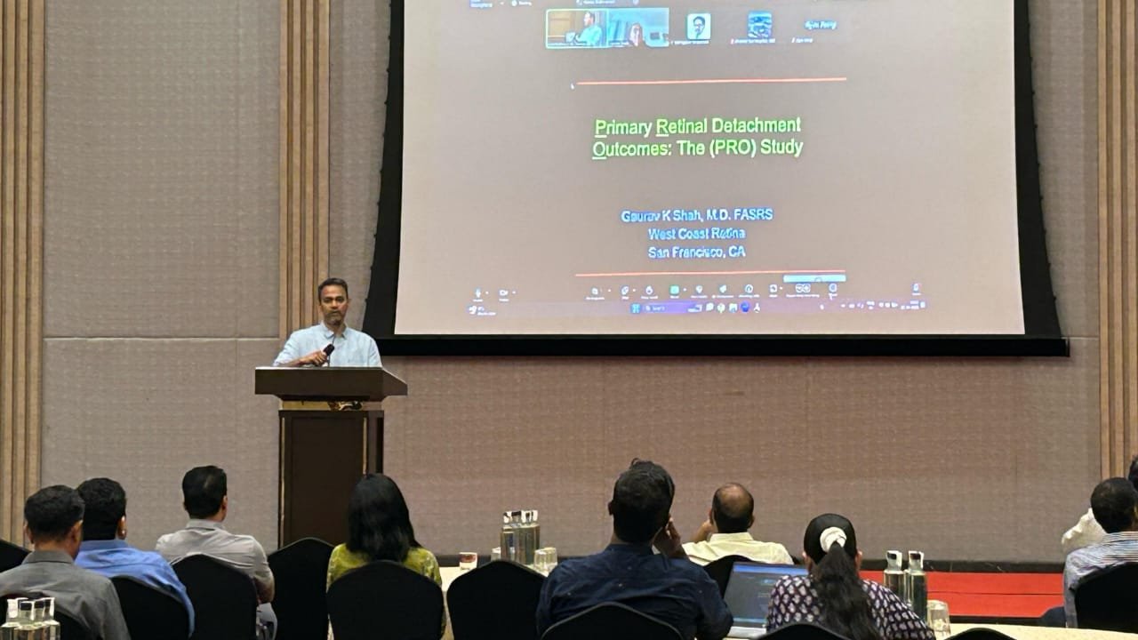
### 9. SALWEEN: 1 YEAR DEEP DIVE
*Presenter:* Dr. Vasumathy V, Radhatri Nethralaya
This presentation provided comprehensive one-year longitudinal experience with the Salween retinal imaging system or therapeutic platform. Content included:
– *Platform Overview:*
– *System Description:* Technical specifications of Salween technology (could be ultra-widefield imaging platform, OCT system, surgical visualization system, or other retinal diagnostic/therapeutic device – presentation clarified specific platform)
– *Institutional Acquisition:* Timeline of implementation at Radhatri Nethralaya, training process, and integration into clinical workflow
– *Intended Applications:* Primary clinical indications for which system was acquired
– *One-Year Experience Metrics:*
– *Patient Volume:* Total number of patients examined/treated using Salween platform during first year
– *Procedure Distribution:* Breakdown of applications (diabetic retinopathy screening, ROP evaluation, surgical planning, etc.)
– *Demographic Analysis:* Age distribution, presenting conditions, and referral patterns
– *Clinical Applications and Case Series:*
– *Diabetic Retinopathy:*
– Number of screening examinations performed
– Detection rates for diabetic macular edema, proliferative changes, and peripheral lesions
– Clinical decision impacts (treatment initiation, referral for laser/injection)
– *Retinopathy of Prematurity:*
– Integration into ROP screening program
– Comparison with traditional indirect ophthalmoscopy
– Telemedicine applications for remote screening
– *Retinal Vascular Diseases:*
– Vein occlusions, arterial occlusions, and vasculitis evaluation
– Peripheral pathology detection enhancing diagnosis
– *Surgical Planning:*
– Preoperative assessment for vitrectomy cases
– Documentation of pathology extent and surgical approach determination
– *Other Applications:*
– Uveitis monitoring, inherited retinal diseases, tumor evaluation
– *Advantages Identified:*
– *Enhanced Detection:* Cases where Salween imaging identified pathology missed on conventional examination/imaging
– *Patient Engagement:* Improved patient understanding through visualization of their own pathology
– *Documentation Quality:* Superior image quality for medical records and medico-legal purposes
– *Workflow Efficiency:* Time savings or workflow improvements compared to previous protocols
– *Telemedicine Enablement:* Facilitation of remote consultations and second opinions
– *Challenges and Limitations:*
– *Learning Curve:* Initial difficulties in image acquisition, interpretation, or technical operation
– *Technical Issues:* Equipment malfunctions, maintenance requirements, or software challenges encountered
– *Patient Cooperation:* Cases where pediatric age, cognitive impairment, or poor fixation limited image quality
– *Cost Considerations:* Economic analysis of per-examination costs, maintenance expenses
– *Integration Barriers:* Challenges incorporating into existing electronic medical record or workflow
– *Comparative Analysis:*
– *Versus Previous Technology:* Direct comparison with institution’s prior imaging modality on quality, efficiency, diagnostic yield
– *Benchmark Against Literature:* How institutional outcomes compare with published data on Salween or similar platforms
– *Protocol Evolution:*
– *Initial Protocol:* How system was used in first months of implementation
– *Iterative Refinements:* Adjustments made to imaging protocols, patient selection, or workflow based on experience
– *Current Best Practices:* Optimized protocols developed through one year of use
– *Illustrative Cases:*
– *Case 1:* Dramatic example where Salween imaging was clinically pivotal (e.g., peripheral pathology detection preventing blindness)
– *Case 2:* Challenging case demonstrating platform capabilities or limitations
– *Case 3:* Comparison case showing superiority over conventional imaging
– *Quality Metrics and Outcomes:*
– *Diagnostic Accuracy:* Sensitivity/specificity for various pathologies if formal validation performed
– *Treatment Decisions:* Percentage of cases where imaging directly altered management
– *Patient Satisfaction:* Feedback from patient surveys (if collected)
– *Referring Physician Satisfaction:* Feedback from community ophthalmologists utilizing reports
– *Future Directions:*
– *Expanding Applications:* Plans to utilize for additional conditions or populations
– *Research Initiatives:* Ongoing or planned research projects leveraging Salween platform
– *Training Programs:* Educational initiatives to train fellows/residents on platform
– *AI Integration:* Potential for incorporating artificial intelligence analysis tools
– *Return on Investment:*
– *Financial Analysis:* Revenue generation versus costs over first year
– *Clinical Value:* Improvements in patient outcomes or care quality
– *Strategic Value:* Positioning institution as referral center for advanced imaging
– *Recommendations for Others:*
– *Adoption Considerations:* Factors institutions should weigh when considering Salween acquisition
– *Implementation Tips:* Lessons learned to facilitate smooth integration
– *Training Requirements:* Recommended training pathways for operators and interpreting physicians
—–
## EXPERT PANEL DISCUSSION
*Panel Members:*
– Dr. Vasumathy V
– Dr. Jatinder Singh
– Dr. Parag Shah (Chairperson)
– Dr. V.R. Saravanan
– Dr. M. Prabhu Shanker
– Dr. Ashraya Nayaka T.E.
Following the case presentations, the expert panel facilitated interactive discussion addressing:
### Technology Integration and Clinical Translation
– *Ultra-widefield OCT Adoption:* Panel discussion on practical considerations for incorporating ultra-widefield OCT into regional practices, cost-benefit analysis, and appropriate patient selection
– *AI Implementation Barriers:* Challenges to deploying AI-based ROP screening in Indian NICUs including infrastructure requirements, ophthalmologist training needs, and regulatory landscape
– *Imaging Modality Selection:* Framework for choosing appropriate imaging technology (traditional angiography versus OCTA versus widefield) based on clinical question and available resources
### ROP Management Controversies
– *Anti-VEGF versus Laser:* Expert debate on optimal primary treatment selection considering efficacy, safety, and follow-up reliability in South Indian practice context
– *Follow-up Strategies:* Discussion of systems-level interventions to reduce loss to follow-up rates including appointment reminder protocols, transportation assistance, and community health worker integration
– *Atypical Presentations:* Approach to unusual ROP variants including Coats-like response, considering differential diagnosis and tailored management
### Pediatric Vitreoretinal Challenges
– *Diagnostic Dilemmas:* Expert insights on differentiating retinoblastoma from infectious/inflammatory conditions in children presenting with leukocoria or unilateral vision loss
– *Toxocariasis Management:* Panel perspectives on antihelminthic therapy timing, surgical indications, and visual prognosis communication with families
– *Multidisciplinary Collaboration:* Effective models for coordinating care with pediatricians, neonatologists, and infectious disease specialists
### Regional Practice Considerations
– *Resource Optimization:* Strategies for delivering evidence-based care within resource constraints common in South Indian settings
– *Telemedicine Applications:* Expanding access to subspecialty expertise through remote imaging interpretation and teleconsultation
– *Training Initiatives:* Needs assessment for ROP and pediatric retinal disease education among general ophthalmologists and residents
### Emerging Technologies
– *AI Future Directions:* Beyond ROP screening—potential applications in diabetic retinopathy, AMD, and glaucoma screening
– *Portable Imaging Devices:* Smartphone-based and handheld imaging systems enabling bedside NICU examinations
– *Cost Reduction Trends:* Anticipated technology price decreases expanding access to advanced imaging
### Audience Engagement
The panel encouraged active participation from attendees through:
– *Case-based Polling:* Interactive audience response to clinical scenarios presented during discussion
– *Q&A Session:* Open forum for attendees to pose questions about lectures, cases, or general practice issues
– *Experience Sharing:* Invitation for attendees to share relevant cases or experiences from their practices complementing the discussed topics
—–
## VOTE OF THANKS
*Presenter:* Dr. V.R. Saravanan
Dr. Saravanan concluded the scientific session with formal acknowledgments:
– *Distinguished Guest Speaker:* Deep gratitude to Professor Dr. J. Peter Campbell for traveling from Oregon Health & Science University to share cutting-edge expertise in ultra-widefield OCT and AI for ROP, significantly enriching the regional knowledge base and inspiring adoption of innovative technologies
– *Session Chairperson:* Recognition of Dr. Parag Shah for expertly moderating discussions and introducing the keynote lectures with scholarly context
– *Case Presenters:* Appreciation for all speakers (Dr. Saurab Kapase, Dr. Bidisha P, Dr. Anjali Raju, Dr. Kshitij Donimath, Dr. Vasumathy V) for their excellent case presentations demonstrating clinical expertise and contributing to collective learning through challenging scenarios
– *Expert Panel Members:* Thanks to the distinguished panel (Dr. Vasumathy V, Dr. Jatinder Singh, Dr. Parag Shah, Dr. M. Prabhu Shanker, Dr. Ashraya Nayaka T.E.) for providing valuable insights, facilitating robust discussions, and addressing audience queries
– *Organizing Committee:* Recognition of the logistical coordination, program planning, and execution ensuring a smooth and productive scientific session
– *Participating Institutions:* Acknowledgment of institutional support from Aravind Eye Hospital, The Eye Foundation, and Radhatri Nethralaya, whose faculty contributed significantly to the program
– *Attendees and Delegates:* Gratitude to all ophthalmologists who attended despite busy clinical schedules, contributing their active participation and making the session scientifically vibrant
– *Venue Partners:* Appreciation to Zone by the Park for excellent hospitality, facilities, and technical support enabling a professional meeting environment
Dr. Saravanan emphasized that sessions like these strengthen the regional ophthalmology community, foster collaboration, and ultimately improve patient care through knowledge sharing and adoption of evidence-based innovations.
—–
## LEARNING OUTCOMES ACHIEVED
Participants achieved:
1. *Advanced Imaging Expertise:* Comprehensive understanding of ultra-widefield OCT technology, clinical applications in vitreoretinal surgery, and integration into diagnostic algorithms
1. *AI Knowledge:* Insights into artificial intelligence development for ROP screening, performance characteristics, implementation strategies, and future potential across ophthalmology
1. *ROP Management Proficiency:* Enhanced decision-making for treatment selection (anti-VEGF versus laser), recognition of atypical presentations, and strategies to ensure adequate follow-up adherence
1. *Pediatric Diagnostic Skills:* Improved ability to evaluate children with unilateral vision loss, differentiate serious conditions (retinoblastoma versus infectious/inflammatory), and manage ocular toxocariasis
1. *Non-invasive Imaging Utilization:* Knowledge of OCTA and other dye-free imaging modalities, appropriate clinical applications, and interpretation differences from traditional angiography
1. *Technology Assessment:* Framework for evaluating new diagnostic platforms through longitudinal experience sharing (Salween case study)
1. *Evidence-Based Practice:* Integration of contemporary literature and international expertise into regional clinical practice
—–
## CONTINUING MEDICAL EDUCATION (CME)
This scientific program contributed toward ongoing professional development requirements for ophthalmologists. Participants were advised to maintain attendance documentation for CME credit applications as per Medical Council of India and state medical council regulations.
—–
*Prepared by:* [Organizing Committee Secretary]
*Distribution:* All registered participants, contributing institutions, and CME accreditation bodies
—–
These minutes document the proceedings of the Coimbatore Retina Meet held on March 18, 2026, at Zone by the Park, Coimbatore.